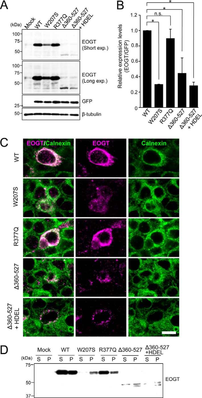
FIGURE 6.

Expression and subcellular localizations of EOGT mutants. A, immunoblot analysis of cell lysates from HEK293T cells transiently expressing wild-type or mutant forms of EOGT. The pSegtag2/EOGT-IRES-GFP constructs allow monitoring of the level of EOGT expression via GFP expression. β-Tubulin was used as a loading control. B, quantification of the EOGT expression level normalized to that of GFP (n = 3; results are shown as mean ± S.D.; *, p < 0.01; n.s., nonspecific; unpaired Student's t test). C, HEK293T cells were transfected to express wild-type EOGT or the indicated EOGT mutants. Immunostaining was performed using anti-EOGT (magenta) and anti-calnexin (green) antibodies. Bar, 10 μm. D, ultracentrifugation of Triton X-100-solubilized EOGT. Note that wild-type EOGT and EOGTR377Q were recovered in the supernatant fraction (S), whereas the majority of EOGTW207S and EOGTΔ360–527 were found in the pellet (P).
