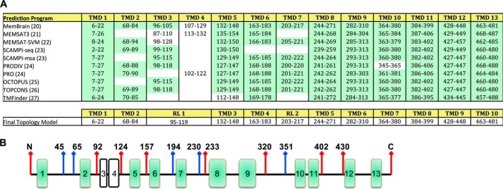
FIGURE 1.
Predicted transmembrane domains for human Hhat. A, TMDs predicted for human Hhat by the indicated programs (top panel). TMDs predicted with high consistency by most programs are highlighted in green. Bottom panel, final topology model of Hhat. RL, re-entrant loop. B, schematic representation of FLAG epitope insert (red arrows) and HA epitope insert (blue arrows) constructs used to map the topology of Hhat.

