FIGURE 5.
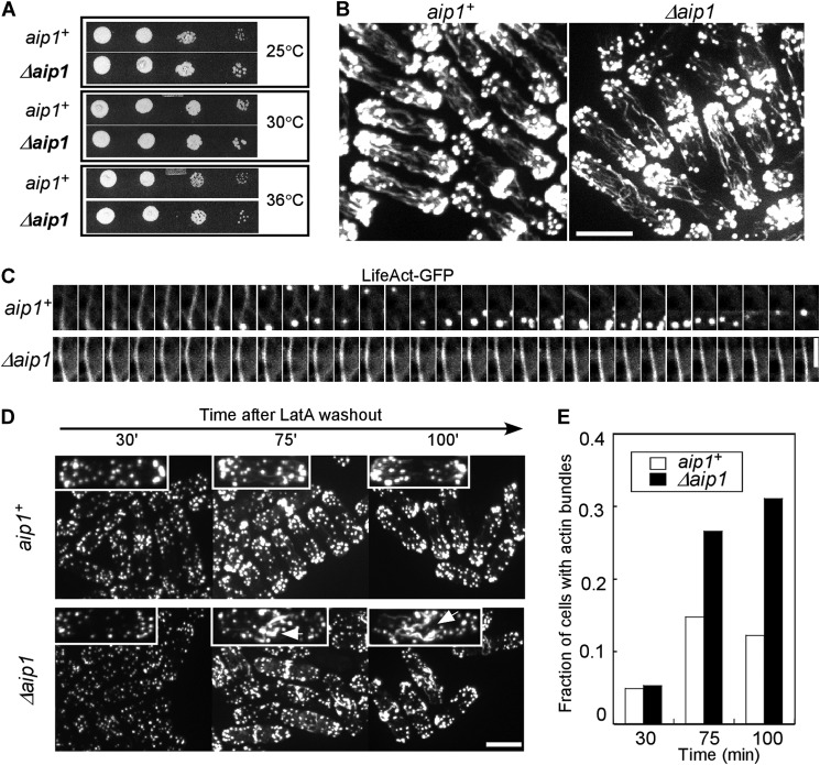
Effects of aip1 deletion on fission yeast. A, serial of dilutions of wild type (aip1+) and deletion mutants (Δaip1) grown at three different temperatures on YE5S plates. B, wild type and Δaip1 cells fixed and stained with Bodipy-phallacidin are indistinguishable in spinning disk confocal fluorescence micrographs. Bar, 5 μm. C, time lapse at 2 s interval of confocal fluorescence micrographs of a single plane of actin cables in a wild type cell and a Δaip1 cell expressing LifeAct-mGFP as a marker for actin filaments. Bar, 2 μm. D, fluorescence micrographs of the time course of the recovery of wild type and Δaip1 cells from treatment with 10 μm LatA for 15 min. Cells were stained with Bodipy-phallacidin 30, 75, and 100 min after washing out LatA. One representative cell is enlarged for each time point. Arrows, thick actin bundles. Bar, 5 μm. E, the histogram shows the fraction of wild type and Δaip1 cells (n > 100) with thick actin bundles at times after LatA washout.

