
FIGURE 6.
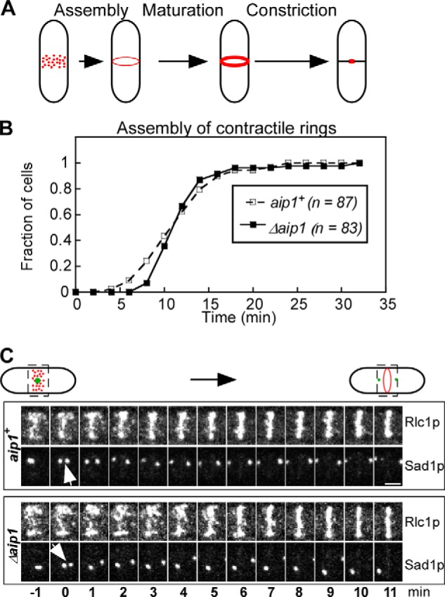
Analysis of contractile ring assembly in wild type and Δaip1 cells expressing Rlc1p-tdTomato and Sad1p-mGFP, related to Table 3. A, cartoon representation of three stages of ring assembly in fission yeast: assembly of the contractile ring from nodes (red dots), maturation of the ring, and constriction. B, outcomes plot of the accumulation of cells with a complete contractile ring over time after the separation of SPBs (time 0). Open squares, wild type cells; filled squares, Δaip1 cells. The difference between the two plots is not statistically significant (p > 0.1), according to a log rank test. C, time series of fluorescence micrographs (maximum intensity projections of Z-series of whole cells) of the division plane (rectangular areas outlined by dashed lines) in a wild type (aip1+) and Δaip1 cell during the assembly of contractile rings. Bar, 2 μm. Numbers are times in min. Arrows point to the separation of SPBs at time 0.