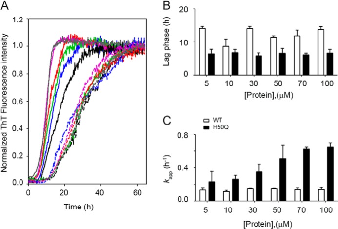
FIGURE 2.

Kinetics of fibrillogenesis. A, normalized fluorescence data of WT (dashed lines) and H50Q (solid lines) α-Syn at six different concentrations: 100 μm (gray), 70 μm (purple), 50 μm (red), 30 μm (green), 10 μm (blue), and 5 μm (black), respectively. B, effect of initial protein concentration on the duration of the lag phase in the aggregation of WT (open bars) and H50Q α-Syn (solid bars), respectively. C, dependence of kapp, apparent growth rate of fibrils, over initial monomer concentration for WT and H50Q α-Syn. The bars represent means ± S.D. of four independent experiments.
