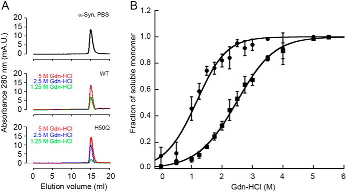
FIGURE 4.

Thermodynamic stability of in vitro fibrils formed by WT and H50Q α-Syn. A, size exclusion profile of ultracentrifuged samples of WT and H50Q α-Syn fibrils after denaturation with guanidine HCl. Representative curves at 5, 2.5, and 1.25 m guanidine HCl, respectively, show a single peak eluting at the same retention time as the native monomer (either WT or H50Q α-Syn) in PBS. Both the two isoforms show the same pattern when applied to a Superdex 200 column equilibrated and eluted with PBS at 0.5 ml/min. mA.U., milli absorbance units. B, the proportion of monomer released from WT (circles) and H50Q (squares) α-Syn fibrils over the total protein concentration at increasing guanidine HCl (Gdn-HCl) concentrations was analyzed with Equation 1 following the linear polymerization model as described under “Experimental Procedures.” Curves shown as mean (S.D.) of three independent experiments.
