
FIGURE 7.
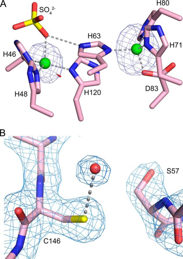
A, identification of zinc ions in the as-isolated, metal-bound C57S hSOD1 structure (PDB entry 4MCM) using x-rays tuned to the copper and zinc absorption edges. Subunit A is shown, which is representative of all 12 SOD1 subunits in the asymmetric unit. The copper-binding site is on the left, and the zinc-binding site is on the right. Zinc ions are shown as green spheres, and a sulfate ion coordinated to the zinc is shown as yellow and red sticks. X-rays tuned to the copper edge do not promote anomalous scattering by zinc and thereby act as the discriminator between the two metal ions. The small amount of electron density shown in red, near the metal in the copper site, comes from the anomalous scattering by copper calculated using data from the copper edge. The electron density shown in blue in both metal-binding sites comes from the anomalous scattering by zinc calculated using data from the zinc edge. The copper edge and zinc edge anomalous difference Fourier electron density maps are both contoured at 5 σ. B, apo C57S hSOD1 (PDB entry 4MCN) lacks the intrasubunit disulfide bond. σ-A weighed electron density with coefficients 2mFo − DFc (63) superimposed on the refined structure of C57S SOD1 in subunit A. The electron density map is contoured at 1.3 σ. The rotation of Ser-57 away from Cys-146 is clearly shown; the distance between the sulfur of Cys-146 and the oxygen of Ser-57 is 5 Å. The red sphere coordinated to Cys-146 is a water molecule.