FIGURE 7.
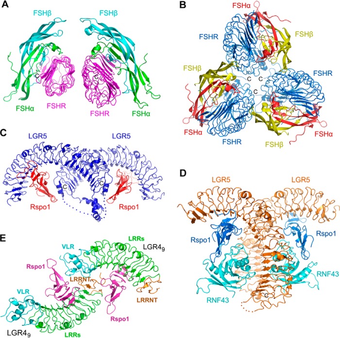
Oligomizeration of LGRs in crystals. A, dimerization of FSH-FSHR complex in an asymmetric unit (PDB code 1XWD). The LRRCT/hinge-truncated ectodomain of FSHR is colored in purple. The α- and β-chain of FSH are colored in green and cyan, respectively. B, trimerization of FSH-FSHR complex in an asymmetric unit (PDB code 4AY9). The entire ectodomain of FSHR is colored in blue. The α- and β-chains of FSH are colored in red and yellow, respectively. C, dimerization of the LGR5-Rspo1 binary complex in an asymmetric unit (PDB code 4BSR). LGR5 and Rspo1 are colored in blue and red, respectively. The disordered loops in the LRRCT region of LGR5 are shown as dashed lines. D, dimerization of the LGR5-Rspo1-RNF43 ternary complex in an asymmetric unit (PDB code 4KNG). LGR5, Rspo1, and RNF43 are colored in orange, blue, and cyan, respectively. E, two LGR49-Rspo1 copies in an asymmetric unit in our complex structure. The two LGR4-Rspo1 copies interact with each other via the engineered VLR module and the LRRNT region.

