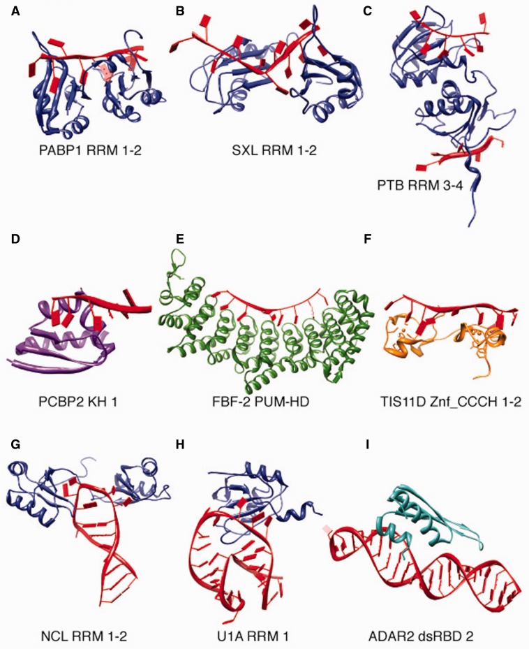
Figure 1:
RNA-binding domains use a variety of strategies for binding RNA. (A–C), Different arrangements of two RRM domains. (A) RRMs 1–2 of PABP1 are arranged to form a flat RNA-binding surface (PDB ID: ICVJ). (B) RRMs 1–2 of SXL form an RNA-binding cleft (1B7F). (C) RRMs 3–4 of PTB are arranged back to back (2ADC). (D–F) Examples of other RNA-binding domains. (D) KH domain 1 of PCBP2 forms an RNA-binding cleft (2PY9). (E) The Puf repeats of the FBF-2 PUM-HD form a concave RNA-binding surface (3K62). (F) The two CCCH zinc fingers of TIS11D/ZFP36L2 (1RGO). (G–I) RBPs binding to structured RNA. (G) Hairpin loop recognition by RRMs 1–2 of Nucleolin (1RKJ). (H) Bulge loop recognition by RRM 1 of U1A/SNRPA (1AUD). (I) dsRNA binding by the dsRBD domain of ADAR2 (2L2K).

