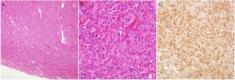
Fig. 3.
(A) The tumor was a circumscribed mass and had many variable sized vessels (H&E, ×100). (B) The tumor was composed of high cellular spindle cells having irregular patterns and moderate cytologic atypia. Mitosis was occasionally seen (H&E, ×400). (C) The tumor cells were reactive for CD34 (×200).

