
Figure 2.
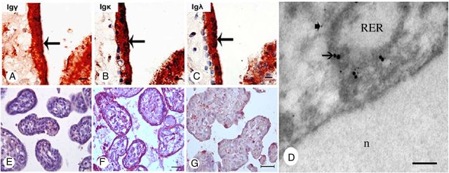
IgG immunoreactivity present in the trophoblasts of the human placenta. (A–C) Immunoreactivity of IgG γ, IgG κ and IgG λ detected in the trophoblasts (arrows). (D) Double electron microscopy ISH and immunostaining to demonstrate co-localization of IgG immunoreactivity and IgG mRNA on the membrane of RER of trophoblast with 15 nm (arrows) and 5 nm (arrow-heads) colloidal gold particles. (E) Symmetric IgG did not react to any cell of the placenta. (F) aIgG isolated from the placenta reacted positively to the trophoblasts and endothelial cells of the placenta. (G) Positive signal disappeared with pre-absorbed IgG. The nuclei were stained blue with hematoxylin. A–C, scale bar = 10 μm; D, scale bar = 100 nm; E–G, scale bar = 10 μm.