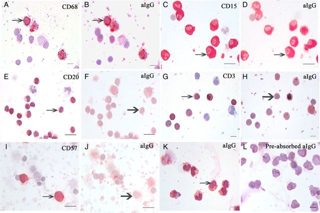
Figure 6.
Reaction of placenta-extracted aIgG with different leukocyte types demonstrated with the stain-decolorize-stain method and the pre-absorption test. (A and B) aIgG reacted to CD68-positive monocytes (arrows point to the same monocyte). (C and D) aIgG reacted to CD15-positive neutrophils (arrows point to the same neutrophils). (E and F) CD20-positive B lymphocytes were negative for aIgG (thick arrows point to the same B-cell). (G and H) CD3-positive T lymphocytes were negative for aIgG (thick arrows point to the same T-cell). (I and J) CD57-positive natural killer (NK) cells were negative for aIgG (thick arrows point to the same NK cell). (K and L) aIgG-positive neutrophils (K) were abolished when the antibody was pre-incubated with IgG molecule in a pre-absorption test on a different slide of the same neutrophil preparation (L). The reactions occurred with leukocytes from the same and different individuals. Symmetric IgG did not react to any leukocyte (not shown). Scale bar, 10 μm.

