
Fig. 2.
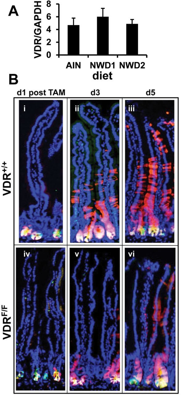
The VDR in intestinal stem cell function. (A) GFP marked cells were isolated by FACS from single cell suspensions of purified intestinal crypts of Lgr5 GFP+ mice fed AIN76A, NWD1 or NWD2 diet from weaning for 6 months. mRNA levels for VDR and for GAPDH were determined by quantitative real-time PCR for the Lgr5 GFP+ cells of three mice in each dietary group (mean ± std) with each sample assayed in quadruplicate; (B) Lgr5 GFP+ , Rosa26 RFP+ , VDR flox/flox or +/+ mice, nine mice per group were fed AIN76A diet for 3 months, given a single tamoxifen injection and three mice of each group killed 1, 3 or 5 days thereafter. Frozen sections of the intestine were stained with DAPI.