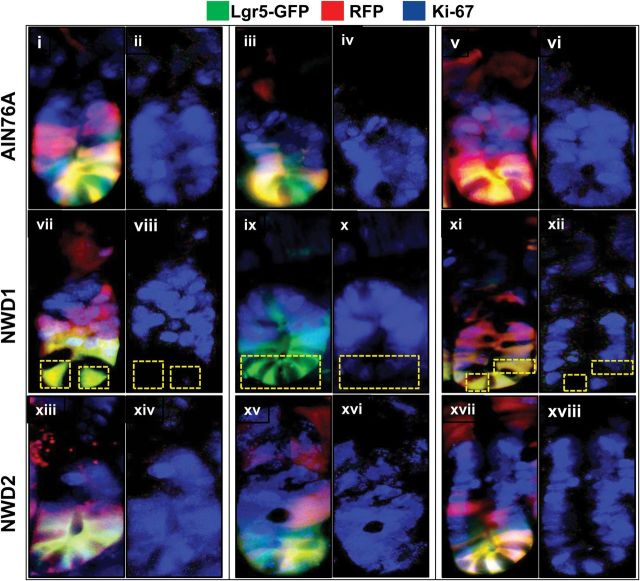
Fig. 4.
Ki67 postive cells at the crypt base in mice fed different diets. Lgr5 GFP+ , Rosa26 RFP+ mice were from the experiment shown in Figure 1B: mice were fed AIN76A, NWD1 or NWD2 diets from weaning, given a single injection of tamoxifen at 3 months and then killed 3 days later, three mice per dietary group. Frozen sections of the intestine were stained with antibody to Ki67 (rabbit anti-mouse, Novus Biologicals, 1:200) detected with a secondary goat anti-rabbit Ab conjugated to Alexa Fluor 350 (InVitrogen, 1:200). The crypts were photographed separately for green, red and blue emission, representing the GFP and RFP expressed in Lgr5+ cells and their progeny, and the expression of Ki67, respectively. Images at the three different wavelengths for each crypt shown were overlayed; shown next to each of these crypts is the isolated blue fluorescence, indicating expression of Ki67.

