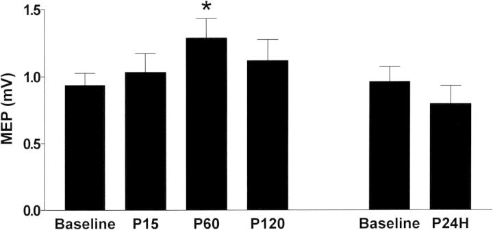
Figure 2.
Effects of left parietal ccPAS on MEPs of left M1HAND. MEPs by left M1 TMS were recorded from right FDI muscle and were averaged from all subjects. There was a significant effect of Time in the corresponding repeated-measures ANOVA; the post hoc analysis showed a significant increase in MEP amplitude at P60 relative to the baseline, indicating left parietal ccPAS enhanced the cortical excitability of left M1. *P < 0.05.

