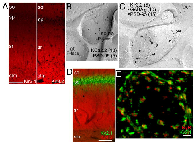
Figure 3. K+ channel localization in dendrites and somata.
A. LM-IF labeling for Kir3.1 (left) and Kir3.2 (right) in rat CA1. From (Kirizs et al., 2014). Scale bar: 100 μm. B. EM-IG labeling for KCa2.2 (large particles, 10 nm) and PSD-95 (small particles, 5 nm, at arrow) showing extrasynaptic KCa2.2 within a dendritic spine (sp) at an excitatory synapse (at: axon terminal) in a SDS-FRL sample from mouse CA1. Scale bar: 0.2 μm. From (Ballesteros-Merino et al., 2012). C. EM-IG labeling of Kir3.2, GABA-B1 receptors, and PSD-95 in a SDS-FRL sample from rat CA1 showing colocalization of Kir3.2, GABA-B1 receptors in the extrasynaptic membrane of a dendritic spine (s) emerging from a dendrite (Den). Scale bar: 0.2 μm. From (Kulik et al., 2006). D. Double LM-IF labeling for Kv4.2 (red) and Kv2.1 (green) in rat CA1 showing precise compartmentalization of these KChs across different hippocampal strata. Scale bar: 100 μm. E. Super-resolution (SIM) image of a double label LM-IF of Kv2.1 (green) and ryanodine receptor (red) in a mouse striatal medium spiny neuron. Scale bar: 2 μm. From (Mandikian et al., 2014). Anatomical labels for panels A, D as in Figure 2.

