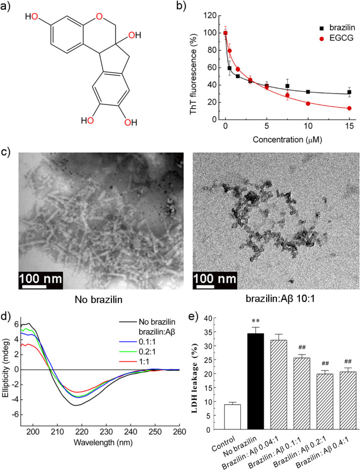
Figure 1. Inhibition of Aβ42 fibrillogenesis and reduction of Aβ42 cytotoxicity by brazilin.
(a) Structural formula of brazilin. (b) ThT fluorescence of Aβ42 (25 μM) aggregates after incubation with various concentrations of brazilin or EGCG for 24 h. See method section for more details. The ThT fluorescence of Aβ42 aggregates without an inhibitor was defined as 100%. The inhibitory potency of brazilin represents a dose-dependent manner with an IC50 of 1.5 ± 0.3 μM, comparing with 2.4 ± 0.4 μM of EGCG. (c) TEM images of Aβ42 in the absence (left) and presence (right) of brazilin (brazilin to Aβ42 ratio, 10:1) after 10 h incubation. (d) The far-UV circular dichroism spectra of Aβ42 incubated for 24 h in the absence and presence of different concentrations of brazilin. (e) Inhibitory effect of brazilin on the cytotoxicity induced by Aβ42 aggregation. Aβ42 monomer (25 μM) was co-incubated at 37°C for 24 h with or without brazilin, and then added to SH-SY5Y cells. After 48 h treatment, cytotoxicity was evaluated using LDH leakage assay. All values represent means ± s.d. (n = 3). ** p < 0.01, compared to control group. ## p < 0.01, compared to Aβ42-treated group.

