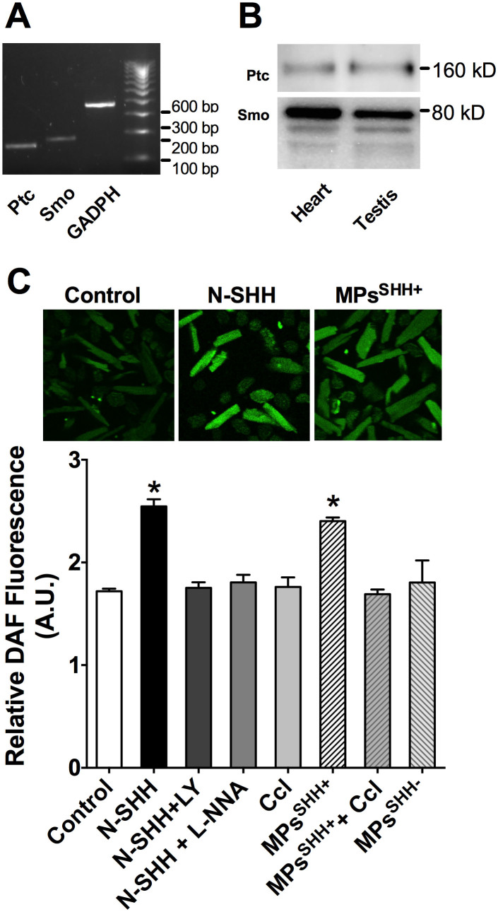
Figure 2. SHH mediates NO production in ventricular cardiomyocytes.
(A) Representative PCR gel showing the expression of Patched (Ptc) and Smoothened (Smo) and reference Glyceraldehyde 3-phosphate dehydrogenase (GAPDH) mRNAs in rat cardiomyocytes. (B) Smo and Ptc expression was revealed by Western blotting in cardiomyocytes and in testis used as reference tissue. These representative images correspond to extracted portion of membranes after transfer of the same gel and exposed respectively with anti- Smo and Ptc primary antibodies. Each band corresponds to adjacent wells of the gel. (C) Representative confocal images illustrating the increase in NO production after incubation of control cardiomyocytes with the recombinant SHH protein (N-SHH) or microparticles harboring the SHH protein (MPsSHH+) for 4 h (upper panel). DAF fluorescence level (lower panel) was quantified in control (n = 495 cells), after incubation with N-SHH (n = 365 cells), and after co-incubation with phosphoinositide-3 kinase inhibitor, LY294002 (LY, 25 μM, n = 98 cells), NOS inhibitor, NΩ-nitro-L-arginine (L-NNA, 100 μM, n = 98 cells). DAF fluorescence level was also quantified after incubation with MPsSHH+ (n = 382 cells) and after co-incubation with the SHH pathway inhibitor cyclopamine (MPsSHH+-Ccl, 30 μM, n = 137 cells). Cyclopamine alone had no effect on basal levels of NO (Ccl, 30 μM, n = 68 cells). Incubation with MPs not carrying the SHH (MPsSHH-) did not enhance NO formation (n = 23 cells). Data are mean ± SEM; *, p < 0.05 compared to control.

