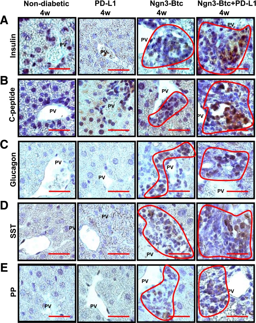
Figure 2.
Ngn3-Btc+PD-L1 induces neo-islets in the periportal regions of the liver. A–E: Representative sections of Ngn3-Btc+PD-L1–treated diabetic NOD mouse liver stained by immunohistochemistry for insulin (A); C-peptide (B); glucagon (C); somatostatin (SST) (D); and pancreatic polypeptide (PP) (E) at 4 weeks (4w) after diabetes onset or treatment. Scale bar = 20 μm. Periportal clusters of cells were only occasionally seen in the PD-L1 group and are shown in A and B to contrast their staining with that of the Ngn3-Btc and Ngn3-Btc+PD-L1 groups. Most of the periportal areas in the PD-L1 group were similar to those shown in C–E. PV, portal vein.

