
Figure 1.
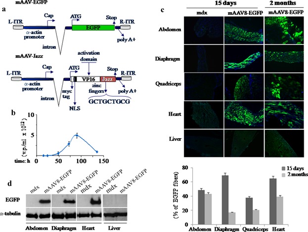
mAAV virus vectors. a: Schematic representation of the engineered mAAV-EGFP and mAAV-Jazz vectors under the control of the muscle-specific human α-actin promoter. b: Time course titration curve obtained by qPCRs performed with growth medium fractions containing viral particles from AAV-293 transfected cells. c, top: EGFP expression in skeletal and cardiac muscles and liver tissues. The mAAV-EGFP-treated and untreated mdx mice were injected intraperitoneally at 5 days of age. Injections were performed with 150 µl of mAAV8-EGFP virus suspension at the concentration of 5 × 10¹² v.p./ml, or with the same volume of saline solution. At different times after injection mice were analyzed and cryostatic sections were examined for direct EGFP fluorescence. Nuclei are stained in blue with Dapi. All the images were taken at 10× magnification. c, bottom: The number of EGFP fibers for each group was obtained by normalizing to the number of total fibers per cross-sectional area. The resulting percentages at the indicate times are reported in a histogram. d: Evaluation of EGFP protein expression by Western blot analysis in skeletal and cardiac muscles and in liver tissues. Five-day-old mdx mice were injected as above and examined 15 days after injection using the polyclonal antibody against EGFP. Detection of α-tubulin was used to normalize the amount of proteins.