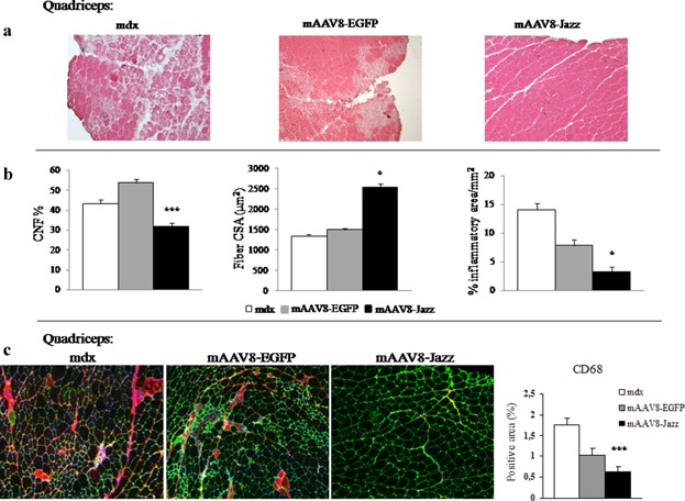
Figure 3.

mAAV8-Jazz ameliorates mdx muscle morphology. a: H&E staining of the quadriceps muscle from 2-month-old untreated, mAAV8-EGFP-treated, and mAAV8-Jazz-treated mdx mice. Reduction in degeneration, necrotic foci, and inflammatory cells is observed in mAAV8-Jazz-treated myofibers (representative sections out of six mice examined in each group). b: Quantification of central nucleation (CNF), cross-sectional area (CSA), and inflammatory infiltrates on H&E stained sections of quadriceps muscle from 2-month-old untreated, mAAV8-EGFP-treated, and mAAV8-Jazz-treated mdx mice (six mice were analyzed in each group; the number of CNFs was obtained by normalizing to the number of total myofibers per CSA, and at least 200 myofibers per section were counted). c: Immunohistochemistry of the quadriceps muscle isolated from 2-month-old untreated, mAAV8-Jazz-treated, and mAAV-EGFP-treated mdx mice (four mice were analyzed in each group). Quantification of macrophage infiltration by staining with CD68 monoclonal antibody (red). The extracellular matrix is counterstained with the anti-laminin polyclonal antibody (green). Nuclei are stained with Dapi (blue; see Materials and Medhods section). Right: Graph shows the quantification of the CD68-positive area (four mice were analyzed in each group). All values are expressed as the mean ± SEM. *P < 0.05 and ***P < 0.001 indicate statistical significance by t-test.
