
Figure 1.
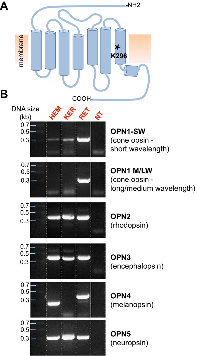
Identification of mRNA corresponding to the retinal-binding site of human opsins in HEMs and KERs. (A) Schematic representation of the topology of human opsins, seven transmembrane domain light-activated receptors that contain in the seventh transmembrane domain a conserved Lys residue (K296 in rhodopsin) required for binding retinal. (B) PCR screen of HEM, KER, retina (RET) or brain (BR) cDNA with primers encompassing the conserved lysine residue (Fig. S1) of cone opsin, short wavelength (OPN1-SW), medium and long wavelength (OPN1-M/LW), rhodopsin (OPN2), encephalopsin (OPN3), melanopsin (OPN4) and neuropsin (OPN5). DNA bands corresponding to OPN1-SW, OPN2, OPN3 and OPN5, but not OPN1-M/LW and OPN4, were amplified from HEMs and KERs. Retina cDNA was used as a positive control for OPN1-SW, OPN1-M/LW, OPN2 and OPN4, and brain cDNA for OPN3 and OPN5. NT, no template. Dotted lines separate the gel lanes, continuous white lines denote different gel lanes joined together.