
Figure 4.
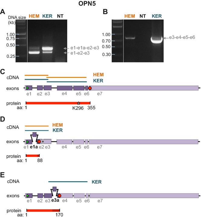
Identification of full length and two truncated splice variants of OPN5 in HEMs and KERs. (A) RT-PCR using HEM or KER RNA and primers based on exon 1 and 3 of OPN5 amplified two bands that corresponded to exons 1–3 (e1-e2-e3, lower band) and to a splice variant containing an additional exon e1a (e1-e1a-e2-e3, upper band). (B) RT-PCR using HEM or KER RNA and primers based on exon 3 and 6 of OPN5 amplified a band that corresponded to exons 3–6 (e3-e4-e5-e6). (C) The lower cDNA bands amplified in (A) and the bands amplified in (B) correspond to exons 1–6 of OPN5, coding for a 355 amino acid peptide containing the retinal binding site K296. (D) The upper cDNA band amplified in (A) contains an additional 89 bp exon (e1a) between exons 1 and 2. The coding sequence of this splice variant translates into an 88 amino acid peptide (Fig. S3A). (E) The cDNA band amplified in B from KERs comprises, in addition to the fragment shown in (C), a splice variant containing an additional 76 bp exon (e3a) between exons 3 and 4 and results in a 170 amino acid peptide (Fig. S3B).