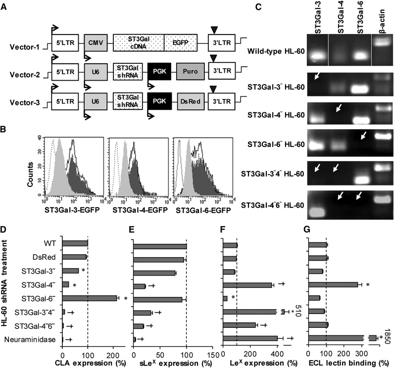
Figure 2.
Effect of α(2,3) sialylT silencing on cell-surface glycan expression. (A) Lentiviral vectors used for shRNA screening and gene silencing. Stable CHO-S cells expressing the ST3Gal-EGFP fusion protein were made using the pCS-CG construct (vector 1). pLKO.1 vectors 2 and 3 carry the shRNA along with either puromycin drug selection (vector 2) or DsRed fluorescent reporter (vector 3). (B) Solid empty histograms in all panels present CHO-S cells stably expressing 1 of the ST3Gal-EGFP fusion proteins. Transduction of these cells with lentivirus carrying shRNA against corresponding ST3Gal variants (gray filled histograms), but not control shRNA (black filled histogram), resulted in a reduction in EGFP signal as measured using flow cytometry. Dashed empty histograms WT CHO-S cells without EGFP fusion protein. Efficient shRNA that reduced EGFP fluorescence by >80% were selected for functional studies. (C) Gel electrophoresis analysis of RT-PCR products demonstrate the absence of target mRNA (white arrow) in single and dual knockdown HL-60 cells. Similar results were obtained using quantitative RT-PCR. (D-G) Flow cytometry measured cell-surface expression of the CLA using mAb HECA-452 (D), sLeX/CD15s epitope using mAb CSLEX-1 (E), Lewis-X/CD15 using mAb HI98 (F), and Galβ1,4GlcNAc/N-acetyllactosamine using ECL lectin (G). All data were normalized with respect to WT HL-60s (dashed line). *P < .05 with respect to all other treatments, †P < .05 with respect to all other treatments except daggers (†) are not different from each other.

