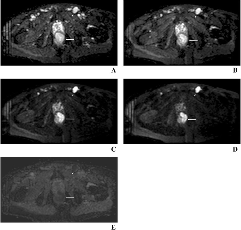
Figure 5.

Anal cancer (arrow) and metastasis in an inguinal lymph node (arrowhead) in the same patient as in Figure 3 . Examination with a 1.5-T pelvic phased array coil. DWI images at b = 0 (A), 50 (B), 500 (C) and 1,000 s/mm2 (D) as well as the corresponding apparent diffusion coefficient (ADC) map (E) at b = 1,000 s/mm2. The ADC value for the tumor and lymph node 0.867 × 10⁻³ and 0.809 × 10⁻³ mm2/s, respectively.
