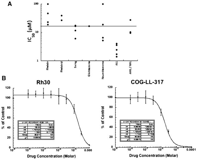
Fig. 1.
PR-104 in vitro activity. A: Relative sensitivity of the cell lines using the IC50 values displayed by histotype. The black line indicates the median IC50 (16.5 μM) for the panel. Cells were exposed to PR-104 for 96 hr under aerobic conditions at concentrations from 10 nM to 100 μM, and viable cells determined by fluorescein diacetate staining. Concentrations of PR-104 that inhibited cell proliferation by 50% (IC50) are plotted for each cell line of each histotype. B: Typical growth inhibition curves for Rh30 and COG-LL-317. Error bars represent standard deviations for each concentration tested.

