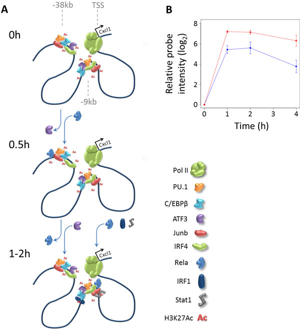
Figure 6.

Cxcl1 enhancer regions illustrating role of transient loss of ATF3 binding. (A) Schematic representation of the genomic region upstream of the Cxcl1 gene, at time points 0h, 0.5h, and 1-2h after stimulation. Two enhancers are shown, one 9kb and one 38kb upstream of the Cxcl1 transcription start site. The H1 enhancer at -38 kb loses ATF3 binding at 0.5h, and gains Rela binding. At the later time points, ATF3 binding is restored, and the enhancer at -9 kb is bound by IRF1, Rela, and STAT1. (B) Average relative probe intensities of the Cxcl1 gene are increased in ATF3 KO compared to WT cells. Average values +/- standard deviation are shown for probe 1457644_s_at (3 replicates), relative to 0h values. Similar plots are shown for two other probes in Supplementary Fig. S5A in Additional file 1.
