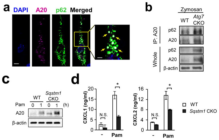
Figure 9. p62 mediates A20 sequestration by autophagy.
(a) Co-localization of A20 and p62 was evaluated by confocal microscopy. NIH-3T3 cells expressing exogenous A20-V5 and p62-HA were stimulated with Pam3CSK4 (100 ng/ml) for 60 min, and A20 (pink) and p62 (green) were detected by V5 and HA antibodies, respectively. White arrows indicate co-localization of A20 and p62. Scale bar = 10 μm. (b) Co-immunoprecipitation of A20 and p62. Peritoneal F4/80hi macrophages were stimulated with Pam3CSK4 (100 ng/ml) for 60 min. Cell lysates were immunoprecipitated with A20 antibody, then p62 was detected with p62 antibody by Western. Results for negative controls using unstimulated cells and control IgG is shown in Suppl. Fig. 15e, f. (c) Expression of A20 in WT and p62 (Sqstm1)-deficient peritoneal F4/80hi macrophages, which were unstimulated or stimulated with Pam3CSK4 (100 ng/ml) for 1 hr. (d) CXCL1 and CXCL2 production by WT and p62 (Sqstm1)-deficient peritoneal F4/80hi macrophages. Macrophages were stimulated with Pam3CSK4 (100 ng/ml) for 24 hrs, and chemokine levels in the culture supernatants were evaluated by ELISA. n=3. Data in all the panels are representative of independent at least two experiments. Error bars represent mean ± SD. ANOVA was used for statistical analysis. N.S.; not significant. *; p < 0.05.

