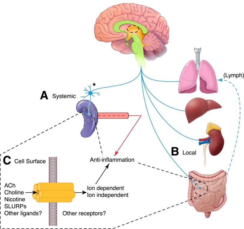
Figure 1. Vagus nerve control of inflammation.
There are 2 anti-inflammatory pathways activated after vagus nerve stimulation. The 1st (A) elicits systemic changes in circulating cytokines and in splenic cells that produce inflammatory and anti-inflammatory cytokines. In this spleen-dependent response, the vagus nerve acts by stimulating a sympathetic response in neurons of the celiac-mesenteric ganglia, which in turn, innervate the spleen where T cells release ACh to alter monocyte homeostasis. In the 2nd pathway (B), a local release of ACh by cholinergic neuronal projections of the vagus nerve targets inflammatory and epithelial cells locally in the lung, liver, kidney, and gut. This pathway, which is spleen independent, regulates local homeostasis and cytokine production in the target tissue, while also gauging cytokines and cells in the lymphatic vasculature (Lymph). In both instances, ACh is released (C) and presumed to act through the α7nAChR, but there is evidence for peptide hormone-like ligands (e.g., SLURP1/2 [26, 27] that can also affect function; see text for further details).

