
Figure 2. Foxp3 expression inhibits Akt activation and Glut1 expression.
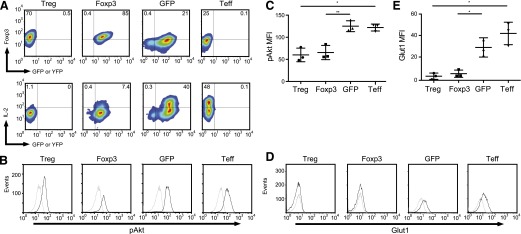
(A, upper) Tregs and Tconv were activated with CD3/CD28 mAb-coated beads and were transduced with lentiviral vectors expressing YFP-2A-Foxp3, GFP, or left untransduced. The correlation between YFP or GFP and Foxp3 was determined by flow cytometry. (Lower) Untransduced Treg, YFP-2A-Foxp3, GFP, and untransduced effector T cells were restimulated with PMA (50 ng/ml) and ionomycin (500 ng/ml) for 3 h, and intracellular IL-2 expression was determined by flow cytometry. (B) Untransduced Treg, YFP-2A-Foxp3, GFP, and untransduced Tconv (as shown in A) were restimulated with CD3/CD28 mAb-coated beads and IL-2 (300 U/ml) for 15 min (black histograms) or left unstimulated for 15 min (gray histograms). Cells were then stained with phospho-Ser473 Akt antibody. (C) Summary of 3 independent experiments as shown in B (*P < 0.01; **P < 0.001). The data were plotted as the mean ± sd. (D) Untransduced Treg, YFP-2A-Foxp3, GFP, and untransduced Tconv (as shown in A) were restimulated with CD3/CD28 mAb-coated beads and IL-2 (300 U/ml) for 20 h (black histograms) or left unstimulated (gray histograms). Cells were then stained with anti-Glut1 antibody. (E) Summary of 3 independent experiments as shown in D (*P < 0.01). The data were plotted as the mean ± sd.