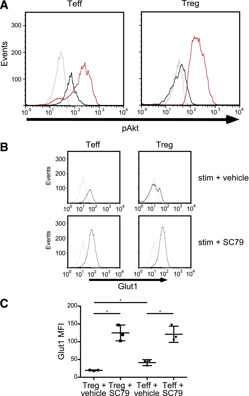
Figure 3. Pharmacologic Akt activation induces Glut1 in Tregs.
(A) Freshly isolated Tconv and Tregs (as shown in Fig. 1A) were stimulated with the Akt agonist SC79 (red histograms), CD3/CD28 mAb-coated beads and 300 U/ml IL-2 (black histograms) or left unstimulated (gray histograms) for 15 min. Cells were then stained with phospho-Ser473 Akt mAb. Data are representative of 3 independent experiments. (B, upper) Freshly isolated Tconv and Tregs (as shown in Fig. 1A) were stimulated with CD3/CD28 mAb-coated beads, IL-2 (300 U/ml), and a vehicle control (black histograms) or left unstimulated (gray histograms) for 20 h. (Lower) Freshly isolated Tconv and Tregs were stimulated with CD3/CD28 mAb-coated beads, IL-2 (300 U/ml), and SC79 (0.5 μg/ml) or left unstimulated for 20 h. Cells were then stained with anti-Glut1 mAb. (C) Summary of 3 independent experiments as shown in B (*P < 0.01). The data were plotted as the mean ± sd.

