
Figure 8.
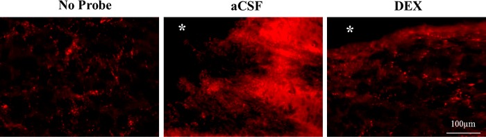
High magnification images (60×) of punctate TH labeling in control tissue (left), tissue near the tracks of probes perfused for 24 h with aCSF (middle) and DEX (right). Punctate TH labeling is diminished near probes perfused with aCSF, and diffuse TH labeling is increased. Control experiments did not indicate nonspecific binding, so the diffuse labeling is presumed to derive from specific binding. Punctate labeling is evident in the DEX image, which does not exhibit diffuse TH labeling. The asterisk near the top of the middle and right-hand images marks a portion of the probe track.