
Figure 1.
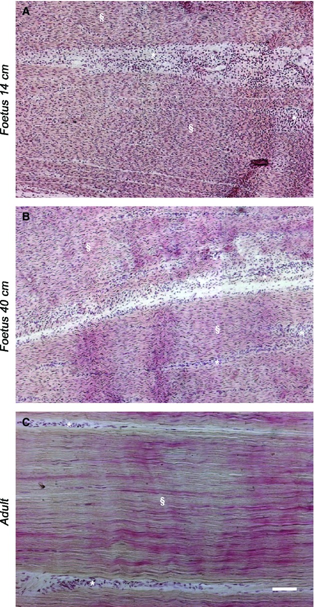
Representative micrographs of HE staining in analyzed tendons. (A) Fourteen-centimeter fetus (mid-pregnancy), (B) 40-cm fetus (late pregnancy) and (C) adult tendon. In fetal tendons (A, B), the endotenon (*) was more developed and thicker (especially in the mid stage fetus one), (A) than that observed in adult tissue (C). Tendon tissue proper (§) is indicated in fetal (A, B) and adult tendons (C). Scale bar: 50 μm.