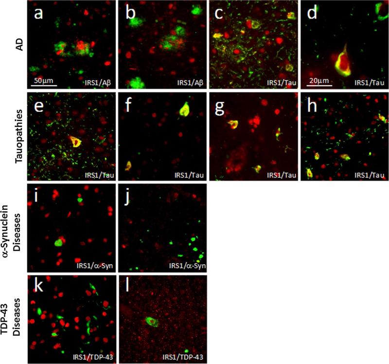
Fig. 3.
Double immunofluorescence images of IRS1-pS616 (red) and neurodegenerative disease lesion proteins (green) in AD (a–d), tauopathies (e–h), α-synucleinopathies (i, j) and TDP43 proteinopathies (k, l). Yellow indicates proteins’ co-localization. a 72-year-old female with AD double labeled for IRS1-pS616 and amyloid-β shows pathological IRS1-pS616 expression in neuronal cytoplasm and neu-rites interspersed among diffuse amyloid-β plaque deposits in mid-frontal cortex. In this and all images except (d), magnification is 400× and scale bar = 50 μm. b 58-year-old male with AD showing IRS1-pS616 immunoreactive neurites enveloped in neuritic amyloid-β plaque and surrounding IRS1-pS616 immunoreactive neurons in mid-frontal cortex; c Same case as in (a) double labeled for IRS1-pS616 and abnormally phosphorylated tau (p-tau) in midfrontal cortex. Note the frequent co-localization (yellow) of perikaryal and neuritic IRS1-pS616 and tau in neurofibrillary tangle-bearing neurons as well as p-tau-immunoreactive neurites; d High magnification (1,000×) image of midfrontal cortex from 68-year-old male with AD shows neurofibrillary tangle that is intensely double labeled for IRS1-pS616 and p-tau neurofibrillary tangle. e 57-year-old male with PiD midfrontal cortex labeled for IRS1-pS616 and p-tau shows double-labeled fibrillar perikaryon of neuron as well as extensive p-tau-immunoreactive neuritic pathology, with some double labeling for IRS1-pS616. f 52-year-old male with CBD shows IRS1-pS616 and p-tau double-labeled neurons. g 68-year-old female with PSP midfrontal cortex double labeled for pS-IRS1 and p-tau shows co-localization in several cells. Such abnormal IRS-1 pS616 expression and p-tau pathology was relatively rare in cortical regions in our sample of PSP cases, prompting us to evaluate IRS-1 pS616 in subcortical regions known to be especially vulnerable in PSP (see text for details). h Pons in 63-year-old female with PSP exhibits extensive IRS1-pS616 and tau co-localization. i Midfrontal cortex in 70-year-old male with PDD immunolabeled for IRS1-pS616 and α-synuclein. Intracellular Lewy inclusions did not co-localize with abnormal IRS1-pS616. j Pons locus coeruleus in 81-year-old male with PD double labeled for IRS-1 pS616 and α-synuclein shows Lewy pathology and no abnormal IRS1-pS616 expression or double labeling. k Midfrontal cortex in 66-year-old female with FTD-TDP immunolabeled for IRS1-pS616 and TDP-43 shows normal-appearing IRS1-pS616 nuclear labeling amidst frequent cytoplasmic and neuritic TDP-43 lesions without co-localization. l Cervical spinal cord from 82-year-old female with ALS shows motor neuron with cytoplasmic TDP-43 surrounding normal IRS1-pS616 labeled nucleus, without co-localization

