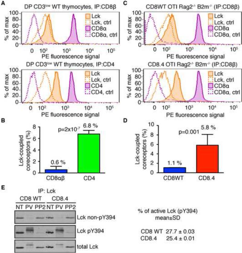
Figure 2. Quantitative determination of Lck coupling to CD4, CD8, and CD8.4 coreceptors.
Cell lysates were incubated with beads coated with antibodies to CD4, CD8β, or isotype controls. Beads were probed with PE-conjugated antibodies to Lck, CD8α, or CD4 and analyzed by flow cytometry. (A-B) Sorted DP CD3low thymocytes from WT mice were analyzed. (C-D) Thymocytes from CD8WT and CD8.4 OTI Rag2−/− β2m−/− mice were analyzed. Representative histograms (A, C) and aggregate data (B, D) (mean ± SD, n = 3-5) are shown. P values were calculated using Student's T test (2 tailed, unequal variance). See also Figure S2. (E) Lck was immunoprecipitated from lysates from nontreated (NT), pervanadate (PV), or 20 μM PP2 treated CD8WT and CD8.4 OTI Rag2−/− β2m−/− thymocytes. Phosphorylation of Lck was analyzed by Western blotting using simultaneous staining with Abs specific for phosphorylated or non-phosphorylated Y394. The membrane was re-probed with Ab to total Lck. Percentage of phosphorylated Lck molecules in resting CD8WT or CD8.4 DP thymocytes was calculated. CD8WT: n=4; CD8.4: n=5.

