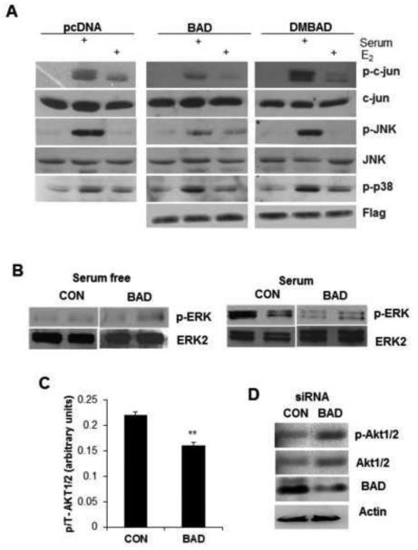
Figure 5. BAD regulates ERK and AKT pathways.
(A) MCF7 cells were transiently transfected with indicated plasmid vectors and were growth arrested with 10nM ICI and 0.1% serum for 48 h prior to stimulation with 5% serum or 10 nM E2. Whole cell lysate (50 μg) of each sample was resolved using SDS-PAGE and immunoblotted with indicated antibodies. (B) After BAD induction for 72 hrs, cells were serum-starved for 24 hrs and stimulated with serum for 1 hr (experiments in duplicate). Lysates from BAD induced and control cells were probed with p-ERK and ERK antibodies. (C) Phospho and total AKT were measured by ELISA and phospho/total (p/T) ratios are shown. Values represent the mean ± S.E. **p<0.01, ***p<0.001 (n=3). (D) Induction of AKT after silencing BAD by siRNA.

