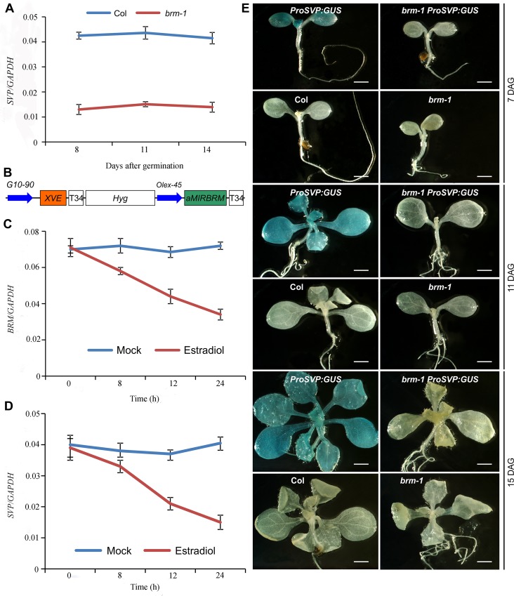
Figure 5. SVP expression is tightly controlled by BRM.
(A) The expression of SVP is drastically decreased in developing brm-1 seedlings compared with that in Col (grown at 22°C) as determined by qRT-PCR. (B) Schematic diagram of the region between the right and left T-DNA borders of the XVE::aMIRBRM construct. The precursor of aMIRBRM was inserted behind a LexA operator sequence fused to the-45 35S minimal promoter (OLexA-45). Other components of the vector were described previously (Curtis and Grossniklaus 2003). (C) BRM expression in 7-old-day XVE::aMIRBRM transgenic seedlings mock treated or treated with 10μm β-estradiol for 0, 8, 12, and 24h, respectively. (D) SVP expression in 7-day-old XVE::aMIRBRM transgenic seedlings mock treated or treated with 10μm β-estradiol for 0, 8, 12, and 24h, respectively. The expression of each gene in A, C, and D was normalized to that of GAPDH. Error bars indicate standard deviations among three technical replicates from one representative experiment. (E) GUS activity patterns of ProSVP:GUS in Col and brm-1 backgrounds in 7, 11, and 14-DAG (days after germination) seedlings. Col and brm-1 were included as negative controls. Scale bar: 0.5 mm.

