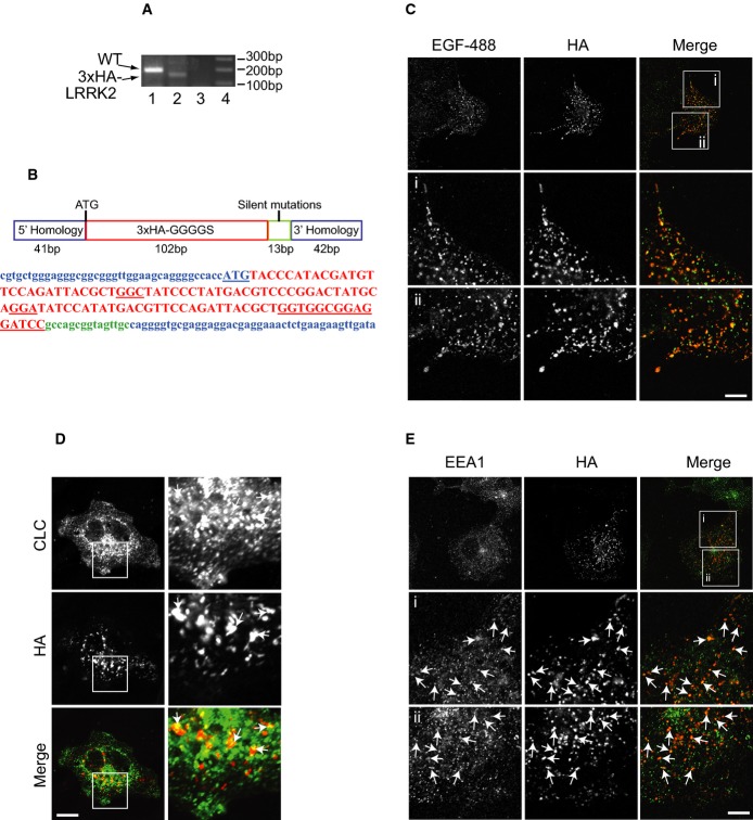
Figure 2. Endogenous genome-edited LRRK2 localizes to endosomes.
- A PCR results of LRRK2-WT from clone E1 (1) using primers that detect endogenous LRRK2. Clone E1 is positive for 3× HA-LRRK2 (2) using a primer pair with the antisense in the 3× HA insert and the sense primer in endogenous LRRK2. (3) Control unedited COS-7 cells using the same primer combination as in (2). (4) 1 kb marker.
- B Schematic diagram of the oligonucleotide used to direct insertion of the 3× HA tag into the 5′ end of the human LRRK2 coding sequence and the corresponding coding sequence in the same colors (the LRRK2 start codon is underlined, as is the GGGGS linker).
- C COS-7 clone E1 cells were serum-starved followed by 20-min incubation with Alexa488-EGF, after which the cells were fixed and processed for immunofluorescence using HA antibody. Scale bar, 10 μm for bottom 6 panels and 25 μm for top 3 panels.
- D, E COS-7 clone E1 cells were fixed and processed for immunofluorescence using HA and CLC (D) or HA and EEA1 (E) antibodies. Scale bar, 10 μm (D, bottom 6 panels in E) and 25 μm (top 3 panels in E).

