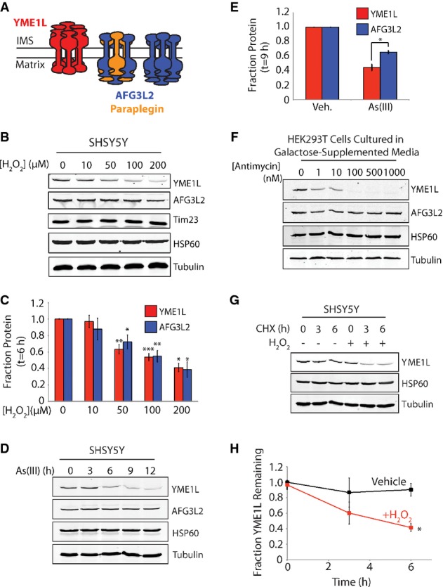
Figure 1. YME1L is a stress-sensitive mitochondrial protease.

- Illustration showing the oligomer assembly and active site orientation of YME1L (red), AFG3L2 (blue), and paraplegin (orange) in the mitochondrial inner membrane.
- Representative immunoblot of lysates prepared from SHSY5Y cells treated with the indicated concentration of H2O2 for 6 h.
- Quantification of YME1L and AFG3L2 from immunoblots as shown in (B). Error bars show SEM for n ≥ 3. ***P-value < 0.001, **P-value < 0.01, *P-value < 0.05.
- Representative immunoblot of lysates prepared from SHSY5Y cells treated with As(III) (50 μM) for the indicated time.
- Quantification of YME1L and AFG3L2 in SHSY5Y cells treated with As(III) (50 μM; 9 h) from immunoblots as shown in (D). Error bars show SEM for n = 4. *P-value < 0.05.
- Immunoblots of lysates prepared from HEK293T cells cultured in galactose-supplemented media treated with the indicated concentration of antimycin A for 6 h.
- Representative immunoblot of lysates prepared from SHSY5Y cells treated as indicated with cycloheximide (CHX; 50 μg ml−1) and H2O2 (100 μM) for the indicated time.
- Quantification of YME1L protein levels from immunoblots as shown in (G). Error bars show SEM for n = 3. *P-value < 0.05.
Source data are available online for this figure.
