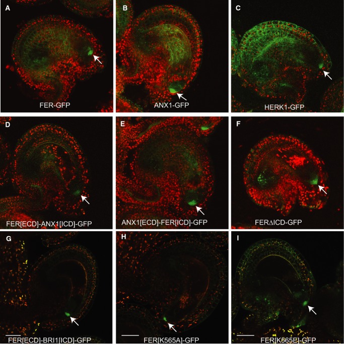
Figure 2. Representative confocal images illustrating that ANX1, HERK1, and domain swap constructs exhibit a FER-like subcellular localization in the filiform apparatus of synergids.
- A FER-GFP is localized to the filiform apparatus of synergids (arrow) and can also be detected at the periphery of sporophytic cells of the ovule.
- B-I All of the GFP fusion constructs show filiform apparatus localization (arrow). While synergid expression and localization to the filiform apparatus was consistent in all lines examined, sporophytic expression in the ovules varied depending on the transgene insertion site. Scale bars are 30 micrometers.
Data information: For all overlayed images, GFP fusion protein signal is shown in green and chlorophyll autofluorescence is shown in red.

