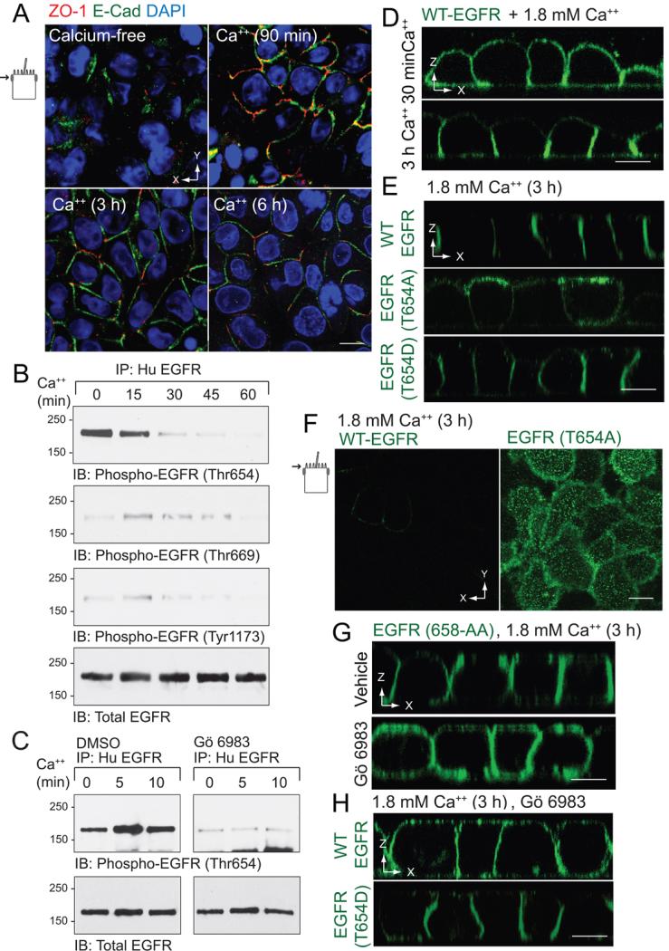
Figure 3. Non-phosphorylatable T654A substitution interferes with BL EGFR polarization in a modified calcium switch model.
Contact naïve MDCK cells with WT-EGFR were plated at confluency on permeable filter supports in calcium-free spinner-modification MEM (S-MEM), re-fed with normal MEM containing 1.8 mM Ca++ 3 h later, and analyzed at times indicated. (A) Confocal images collected in the plane of the Ap membrane (see schematic) from fixed and permeabilized cells stained with antibodies to ZO-1 (red) and E-cadherin (green) and counterstained with DAPI (blue). (B) Human EGFR immune complexes were immunoblotted with phospho-specific EGFR antibodies listed in the figure and an activation-independent EGFR antibody to control for protein loading. (C) Same as (B) except cells were pretreated with DMSO vehicle or the broad spectrum PKC inhibitor Gö 6983 (5 μm) during the calcium-free incubation. (D - H) Cells were fixed, permeabilized, and stained with EGFR1 (green) for confocal imaging. Vertical confocal images of MDCK cells with WT-EGFR 90 min or 3 h after the switch to 1.8 mM Ca++ (D). Vertical confocal images of MDCK cells with WT-EGFR or EGFRs with Thr654 mutations 3 h after the switch to 1.8 mM Ca++ (E). Images collected in plane of the Ap membrane from MDCK cells expressing WT-EGFR or EGFR (T654A) 3 h after the switch to 1.8 mM Ca++ (F). Vertical confocal images of MDCK cells expressing EGFR (658-AA) pre-treated with vehicle (top) or Gö 6983 (5 μm; bottom) 3 h after switch to 1.8 mM Ca++ (G). Vertical confocal images of MDCK cells with WT-EGFR or EGFR (T654D) pre-treated with Gö 6983 3 h after switch to 1.8 mM Ca++ (H). All size bars = 5 μm.

