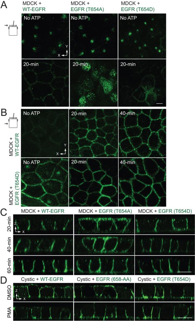
Figure 5. Thr654 regulates BL EGFR delivery in an ATP depletion-repletion renal injury model.
ATP depletion-repletion experiments were carried out exactly as described in Figure 4. (A and B) Horizontal confocal images taken in the plane of the Ap membrane (A) or the middle of the lateral membrane (B) from fixed cells stained with EGFR1 (green) at the end of the 30-min ATP depletion period before ATP repletion (top panels) or following a 20-min incubation in complete media to replenish ATP (bottom panels). (C) Vertical confocal images images from fixed cells stained with EGFR1 (green) at times indicated during the ATP repletion period. (D) Cystic cells derived from the BPK model for ARPKD with stable EGFR expression were incubated with DMSO vehicle or PMA (1 μm) for 30 min, stained with EGFR1 (green) without permeabilization, and analyzed by confocal microscopy. All size bars = 5 μm.

