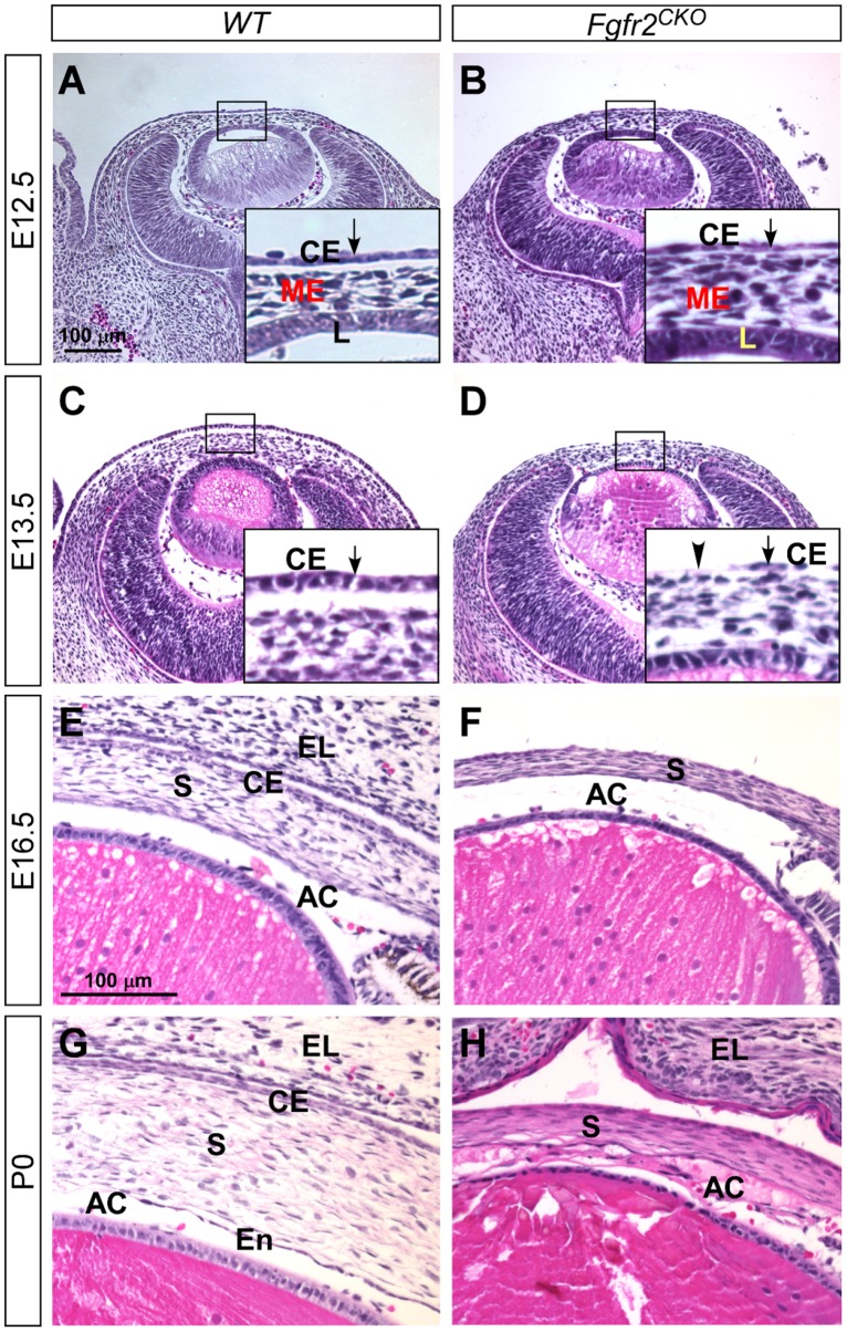
Fig 1. Corneal development (H&E staining) in WT and Fgfr2 CKO eyes.
A, B) At E12.5, ocular mesenchymal cells migrated into the space between the lens (L) and the corneal epithelium (CE, arrow in enlarged inset) in both WT and Fgfr2 CKO eyes. The corneal epithelial layer in Fgfr2 CKO eyes was slightly thinner than that in WT eyes. C, D) At E13.5, the corneal epithelial layer was significantly thinner in Fgfr2 CKO eyes when compared to WT eyes. In some areas, the epithelial cells were absent (arrowhead in D, insert). E, F) At E16.5, corneal stroma (S) was thinner but cells were more densely packed and eosin-staining was intensified in Fgfr2 CKO cornea as compared to WT. Anterior chamber (AC) was formed in both genotypes. As previously reported, eyelid (EL) fusion did not occur in Fgfr2 CKO eyes. G, H) At P0, Fgfr2 CKO mice developed additional defects in the anterior segments, including abnormal accumulation of extra cells and tissues in the anterior chamber (AC), and loss of a distinctive corneal endothelial (En) layer.

