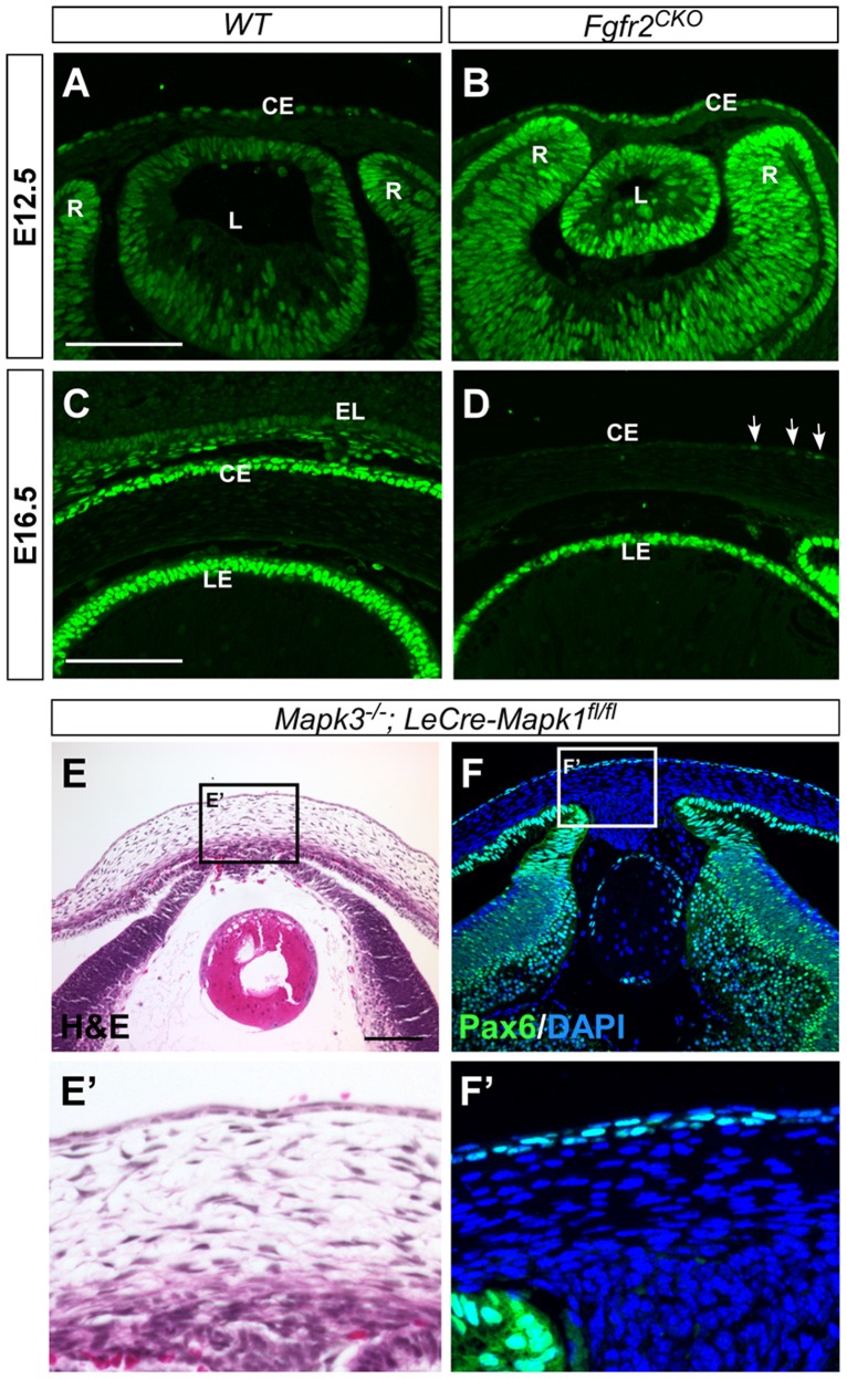
Fig 6. Pax6 immunofluorescence.
A, B) In E12.5 WT and Fgfr2 CKO eyes, Pax6 was expressed in developing corneal epithelial (CE), lens (L) and retinal (R) cells. The expression patterns were similar between WT and Fgfr2 CKO eyes. C, D) At E16.5, Pax6 expression was found in corneal and conjunctival epithelium in WT eyes (C) but was significantly reduced in corneal epithelium of Fgfr2 CKO eye, with a weak signal detected in a few cells (arrows in D). LE, lens epithelium. E, F) Deletion of Mapk1 and Mapk3, encoding for ERK2 and ERK1 respectively, in the surface ectodermal-derived tissues severely affected lens and corneal development (H&E staining in E and E’). However, Pax6 expression appears to be normal in these tissues (F and F’).

