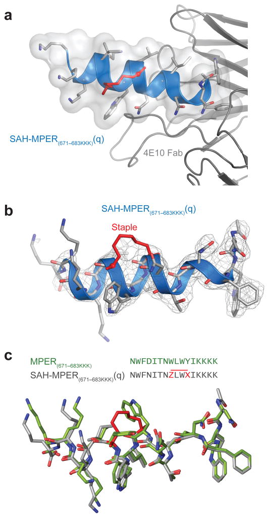
Figure 3. Structural Analysis of the 4E10 Fab–SAH-MPER(671-683KKK)(q) Complex.
(a) Crystal structure of SAH-MPER(671-683KKK)(q) (shown as a blue ribbon and gray transparent van der Waals surface) bound to 4E10 Fab at 2.9 Å resolution. (b) 2Fo-Fc electron density map (1σ level) of the antibody-bound SAH-MPER(671-683KKK)(q) peptide. (c) Superimposition of the native (green, 2FX7)18 and i, i+3-stapled (gray) MPER(671-683KKK) peptides highlights the similarity of antibody-bound structures, aside from the appended C-terminal lysines and the incorporated staple. Z and X represent R3 and S5, respectively.

