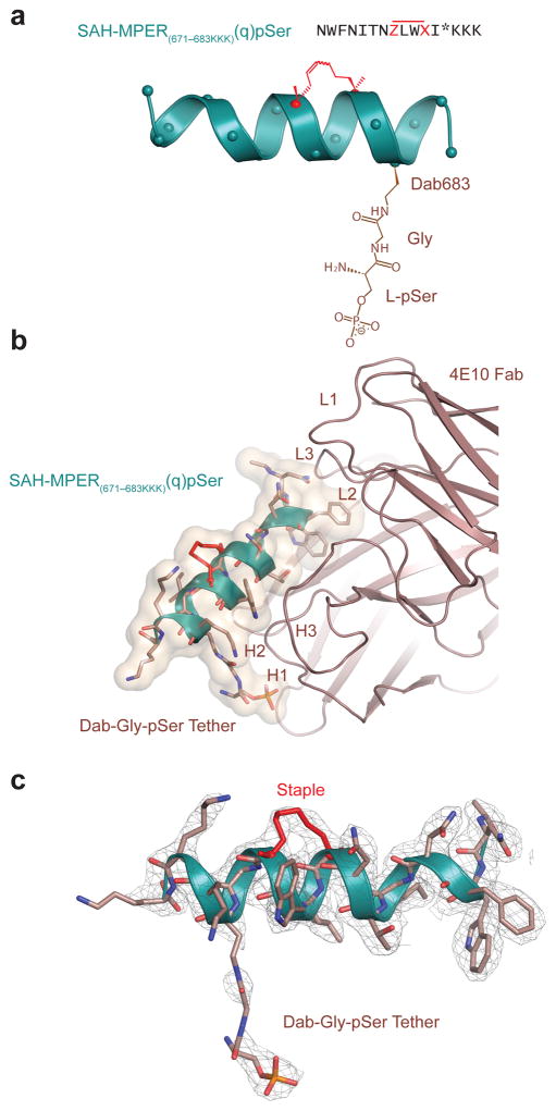
Figure 5. A Phosphate Tether Incorporated into SAH-MPER(671-683KKK)(q) Engages the Putative Lipid Binding Site.
(a) Position 683 of SAH-MPER(671-683KKK)(q) was derivatized with a Dab-Gly-pSer tether (brown) to span the measured 5.5 Å between the primary amine of Lys683 and the phosphate observed at the 4E10 Fab–SAH-MPER(671-683KKK)(q) interface. (b) Crystal structure of the SAH-MPER(671-683KKK)(q)pSer complex (shown as a green ribbon and wheat transparent Van der Waals surface) bound to 4E10 at 2.68 Å resolution. (c) The 2Fo-Fc electron density map (1σ level) of the antibody-bound SAH-MPER(671-683KKK)(q)pSer construct.

