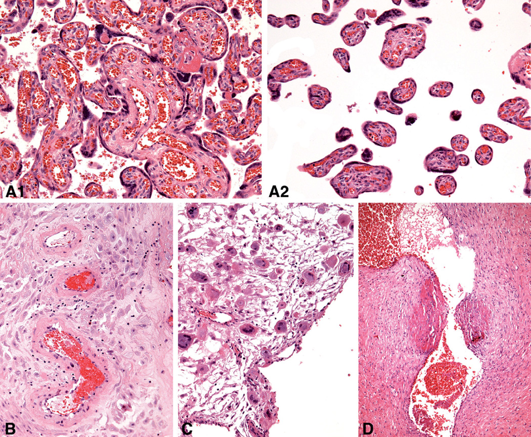
Fig. 1.
Placental lesions for which statistically significant differences were found among 4 groups on hematoxylin-eosin stained sections: A. Uterine pattern of chronic hypoxic placental injury (1 and 2, representative different areas in same placenta), 27 weeks. B. Hypertrophic arteriolopathy in decidua parietalis, 37 weeks. C. Trophoblastic giant cells in decidua basalis, 26 weeks. D. Intimal cushions in a stem vein, 31 weeks. A–C,×20; D,×10

