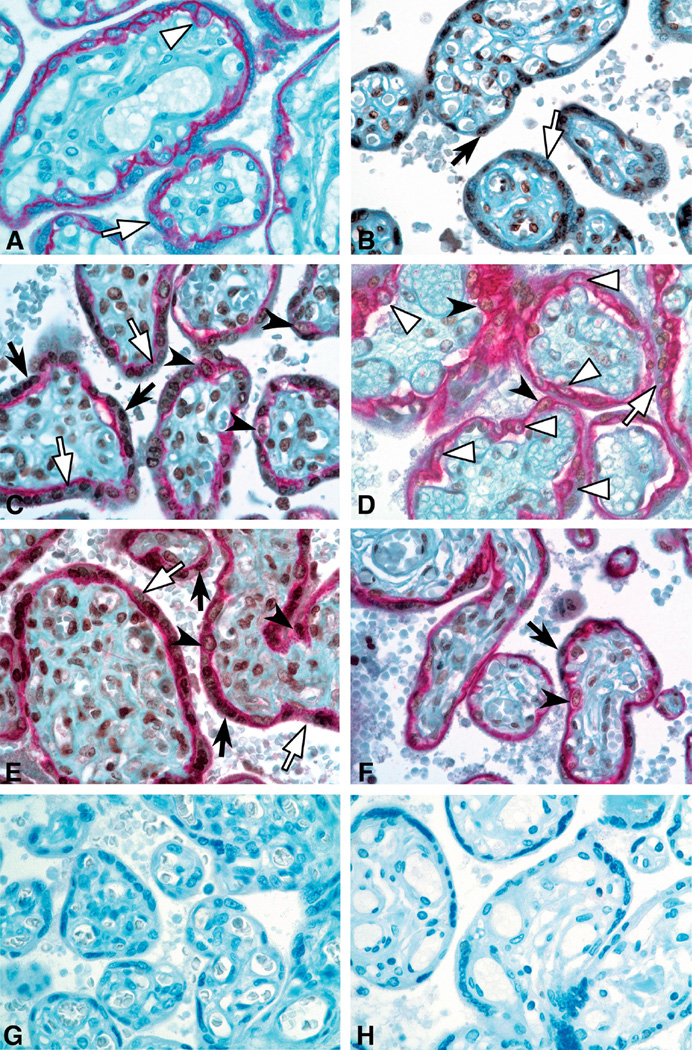
Fig. 2.
Immunohistochemical staining for FOXO1 (brown) and E-cadherin (red) in placental villi (40× magnification). A. E-cadherin antibody only: E-cadherin clearly distinguishes villous CTB, which are mononuclear cells encircled by red membranous staining from STB, which form a mononuclear syncytium with only the basal boundary of STB stained red. B. FOXO1 antibody only: positive cells display brown nuclear staining. Double immunostaining for FOXO1 and E-cadherin in gestational age matched control case (C), mild PE (D), severe PE (E) and FGR (F) placentas at 31 weeks gestation. Empty arrowheads point to FOXO1-negative CTB; solid arrowheads point to FOXO1-positive CTB nuclei; empty arrows point to FOXO1-negative STB nuclei; and solid arrows point to FOXO1-positive STB nuclei. (G, H) Negative control for ecadherin and FOXO1 antibodies, respectively.×40

