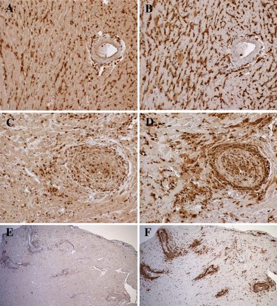
Figure 2.
Male hearts transplanted to female RAG−/− recipients reconstituted with Marilyn CD4 T cells. At 2 weeks, allografts contained diffuse interstitial infiltrates of CD3 T cells (A) and Galectin-3+ macrophages (B) with limited periadventitial involvement of larger arteries. At 6 weeks, the interstitial infiltrates diminished, and large arteries developed adventitial and intimal infiltrates of CD3 T cells (C) and large numbers of macrophages (D). At 10 weeks, the arterial lesions contained decreased numbers of CD3 T cells (E), and increased macrophages (F). Original magnifications 200x (A-D) and 40x (E, F).

