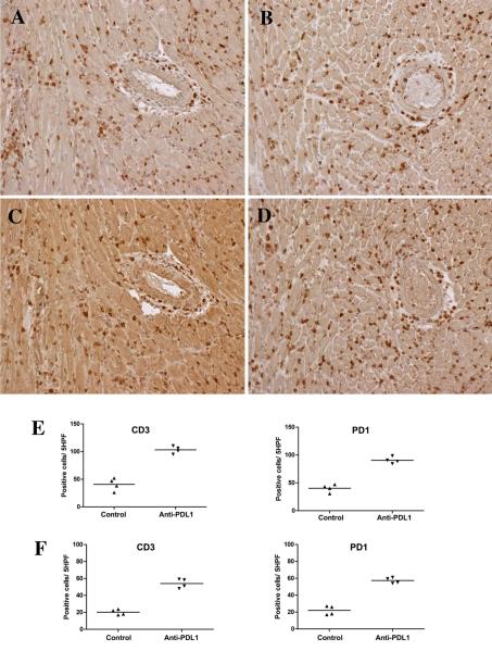
Figure 5.
Immunohistology and cell counts from mice treated with blocking antibodies to PDL1. Administering blocking antibody to PDL1 on days 8, 10 and 12 increased interstitial infiltrates of CD3+ T cells (B) and PD1+ (D) cells compared to controls (A and C, respectively) at 2 weeks. Blocking PDL1 did not increase arterial pathology at 2 weeks (right side of panels A-D). Cell counts per 5 high power fields verified about a 2-fold increase in CD3 and PD1 expressing cells at 2 weeks (E) and 6 weeks (F), but there was an overall decrease in cells from 2 to 6 weeks. Each symbol in the scattergrams represents an individual animal. All differences between control and anti-PDL-1 treated mice were significant <0.05.

